Pages

Friday, April 8, 2011

Breast Infection Causes

Breast Infection Causes

Mastitis (inflammation of breast tissue) is a common benign cause of a breast mass. It is commonly seen in women after childbirth while breastfeeding. These masses are often quite painful. Women who are not breastfeeding can also develop mastitis. In healthy women, mastitis is rare. However, women with diabetes, chronic illness, AIDS, or an impaired immune system may be more susceptible to the development of mastitis.

Bacteria normally found in a baby's mouth or on the nipple can enter the milk ducts through small cracks in the skin of the nipple and can multiply rapidly in the breast milk. This can lead to a superficial small area of inflammation (frequently from streptococcal bacteria) or a deeper walled-off infection or abscess (frequently from staphylococcal bacteria). 

Mild temperature elevations (previously termed milk fever) accompanied by some breast or nipple soreness is usually secondary to engorgement and dehydration immediately (24-72 hours) after delivery and is treated by improved breastfeeding technique. The body temperature should not be above 39°C (102.2°F), nor should the fever persist for longer than about 4-16 hours. This condition may also occur in women who are not breastfeeding and have not completely suppressed lactation yet.
About one to three percent of breastfeeding mothers develop mastitis, usually within the first few weeks after delivery. Most breast infections occur within the first or second month after delivery or at the time of weaning. Typically, the infection is only in one breast. Engorgement and incomplete breast emptying can contribute to the problem and make the symptoms worse.
Chronic mastitis occurs in women who are not breastfeeding. In postmenopausal women, breast infections may be associated with chronic inflammation of the ducts below the nipple. Hormonal changes in the body can cause the milk ducts to become clogged with dead skin cells and debris. These clogged ducts make the breast more prone to bacterial infection. This type of infection tends to come back after treatment with antibiotics.
»»  READMORE...

Tests performed on tumor tissue

Tests performed on tumor tissue

If the pathologist's diagnosis is breast cancer, there are several tests that may be performed on the cancer cells. The results of these tests provide a prognosis and help the oncologist (cancer specialist) guide the patient’s treatment. The most useful of these are HER-2/neu and estrogen and progesterone receptors.

Her-2/neu is an oncogene . It codes for a receptor for a particular growth factor that causes cells to grow. Normal epithelial cells contain two copies of the Her-2/neu gene and produce low levels of the Her-2 protein on the surface of their cells. In about 20-30% of invasive breast cancers, the Her-2/neu gene is amplified and its protein is over-expressed. These tumors are susceptible to treatment that specifically binds to this over-expressed protein. The chemotherapeutic agent Herceptin (Tastuzumab) blocks the protein receptors, inhibiting continued replication and tumor growth. Patients with amplified Her-2/neu gene respond well to Herceptin and have a good prognosis.
Estrogen and progesterone receptor status are important prognostic markers in breast cancer. The higher the percentage of overall cells positive, as well as the greater the intensity, the better the prognosis. Estrogen and/or progesterone receptor positivity in breast cancer cells indicates sensitivity to hormones. The patient may be a good candidate for anti-hormone therapy.
»»  READMORE...

Blood tests

Blood tests
Blood tests may be used to help determine whether or not the tumor is responding to therapy or if it has recurred. Some may be ordered on women who are at a high risk of developing breast cancer to determine whether their risk has a genetic component.
  • CA15-3 (or CA 27.29) is a tumor marker that may be ordered at intervals after treatment to help monitor a patient for breast cancer recurrence. It is not used as a screen for breast cancer but can be used to follow it in some patients once it has been diagnosed.
  • BRCA-1 or BRCA-2 gene mutation – Women who are at high risk because of a personal or strong family history of early onset breast cancer or ovarian cancer can find out if they have a BRCA gene mutation. A mutation in either gene indicates that the patient is at significantly higher lifetime risk (up to 80%) for developing the disease. It is important to remember, however, that only about 5% to 10% of breast cancer cases occur in women with a BRCA gene mutation. Genetic counseling should be considered both before testing takes place and after receiving positive test results.
»»  READMORE...

Wednesday, January 26, 2011

medical health insurance

health VS sickness
insurance benefits 

Health insurance is a type of insurance that protect the availability of funds when an individual or a family member became ill. All the needs of the doctor to have to stay in the hospital with various needs such as, cost of medication in the hospital until all operations are covered by the insurance company. In general, the type of treatment or programs that are available are the benefits of ambulatory (outpatient), benefit-hospital (inpatient), labor benefits and dental benefits.
In general, the benefits of outpatient (Outpatient) incurred by the insurance company is: * GP Consultations * Providing referrals to doctors when necessary spesilis * Consult a medical specialist * The purchase of medicines in accordance with the list of drugs * Pemerikasaan diagnostic support
In the outpatient benefits have maximum limits use of funds each year. Of the total cost of expenditures for outpatient care, in general, in every insurance company requires the insured to pay 20 percent.
Benefits associated with the birth of the child labor, the cost of normal delivery, delivery with additional tools and operating S-caesacea. For there persalian benefit waiting period is usually up to 280 days. Each insurance company has a limit of the waiting period that is different.
As for the benefits of preventive dental care, basic dental care, dental care complex and the installation of dentures. For all treatments insurance companies typically limit the number of total costs that can be used per year. The dental benefits are an obligation for the insured to pay 20 percent of the cost of care (same as outpatient benefits.)
The three benefits of treatment as above, namely outpatient, maternity and dental benefits is an additional option you can take by following the basic program of hospitalization benefits. So you can not just take advantage of outpatient only, labor only, or dental treatment without following the basic program hospitalization benefits.
For the benefit of hospitalization in which the basic benefits that should be taken first, hospitalization benefits coverage in general is almost the same from various insurance companies that exist.The benefits are: * The cost of accommodation space * Fee Non-ICU accommodation * Consult your doctor * Consultation specialists * The cost of surgery, complex, major, intermediate and minor * Other expenses include hospital: drug cost, laboratory tests and diagnostic * Maintain the road 30 days before and after hospitalization * Nursing home * Local ambulance service * Maintain an emergency road accident * Treat dental emergency road accident * Compensation for death
The amount of coverage is very dependent on the program you take, the various insurance companies using the name or type of program is different. For inpatient amount of the premium to be paid annual visits from the cost of accommodation in the hospital room you choose, for example, you choose the cost of accommodation at a cost of Rp. 200 000 then the next following benefit program options specified. For these three additional programs, premium to be paid for outpatient care based on the size of the selected GP consultations, delivery based on the desired cost of natural childbirth and dental care in terms of basic care.
»»  READMORE...

Wednesday, January 19, 2011

Acute renal failure

Acute renal failure
Acute renal failure (ARF) is a rapid loss of renal function due to damage to the kidneys, resulting in retention of nitrogenous (urea and creatinine) and non-nitrogenous waste products that are normally excreted by the kidney. Depending on the severity and duration of the renal dysfunction, this accumulation is accompanied by metabolic disturbances, such as metabolic acidosis (acidification of the blood) and hyperkalaemia (elevated potassium levels), changes in body fluid balance, and effects on many other organ systems. It can be characterised by oliguria or anuria (decrease or cessation of urine production), although nonoliguric ARF may occur. It is a serious disease and treated as a medical emergency.
 
Causes
Renal failure, whether chronic or acute, is usually categorised (as in the flowchart below) according to pre-renal, renal and post-renal causes  
Pre-renal (causes in the blood supply): 
  • hypotension (decreased blood supply), usually from shock or dehydration and fluid loss. 
  • hepatorenal syndrome in which renal perfusion is compromised in liver failure 
  • vascular problems, such as atheroembolic disease and renal vein thrombosis (which can occur as a complication of the nephrotic syndrome) 
Renal (damage to the kidney itself): 
  • infection usually sepsis (systemic inflammation due to infection),rarely of the kidney itself, termed pyelonephritis 
  • toxins or medication (e.g. some NSAIDs, aminoglycoside antibiotics, iodinated contrast, lithium) 
  • rhabdomyolysis (breakdown of muscle tissue) - the resultant release of myoglobin in the blood affects the kidney; it can be caused by injury (especially crush injury and extensive blunt trauma), statins, MDMA (ecstasy) and some other drugs 
  • hemolysis (breakdown of red blood cells) - the hemoglobin damages the tubules; it may be caused by various conditions such as sickle-cell disease, and lupus erythematosus 
  • multiple myeloma, either due to hypercalcemia or "cast nephropathy" (multiple myeloma can also cause chronic renal failure by a different mechanism) 
  • acute glomerulonephritis which may due to a variety of causes, such as anti glomerular basement membrane disease/Goodpasture's syndrome, Wegener's granulomatosis or acute lupus nephritis with systemic lupus erythematosus 
Post-renal (obstructive causes in the urinary tract) due to: 
  • medication interfering with normal bladder emptying. 
  • benign prostatic hypertrophy or prostate cancer. 
  • kidney stones. 
  • due to abdominal malignancy (e.g. ovarian cancer, colorectal cancer). 
  • obstructed urinary catheter. 
 Diagnosis
Renal failure is generally diagnosed either when creatinine or blood urea nitrogen tests are markedly elevated in an ill patient, especially when oliguria is present. Previous measurements of renal function may offer comparison, which is especially important if a patient is known to have chronic renal failure as well. If the cause is not apparent, a large amount of blood tests and examination of a urine specimen is typically performed to elucidate the cause of acute renal failure, medical ultrasonography of the renal tract is essential to rule out obstruction of the urinary tract.
Consensus criteria[1][2] for the diagnosis of ARF are:
  • Risk: serum creatinine increased 1.5 times OR urine production of <0.5 ml/kg body weight for 6 hours .
  • Injury: creatinine 2.0 times OR urine production <0.5 ml/kg for 12 h. 
  • Failure: creatinine 3.0 times OR creatinine >355 μmol/l (with a rise of >44) or urine output below 0.3 ml/kg for 24 h .
  • Loss: persistent ARF or more than four weeks complete loss of kidney function .
Kidney biopsy may be performed in the setting of acute renal failure, to provide a definitive diagnosis and sometimes an idea of the prognosis, unless the cause is clear and appropriate screening investigations are reassuringly negative.
Treatment
Acute renal failure may be reversible if treated promptly and appropriately. The main interventions are monitoring fluid intake and output as closely as possible; insertion of a urinary catheter is useful for monitoring urine output as well as relieving possible bladder outlet obstruction, such as with an enlarged prostate. In the absence of fluid overload, administering intravenous fluids is typically the first step to improve renal function. Fluid administration may be monitored with the use of a central venous catheter to avoid over or under replacement of fluid. If the cause is obstruction of the urinary tract, relief of the obstruction (with a nephrostomy or urinary catheter) may be necessary. Metabolic acidosis and hyperkalemia, the two most serious biochemical manifestations of acute renal failure, may require medical treatment with sodium bicarbonate administration and antihyperkalemic measures, unless dialysis is required.
Should hypotension prove a persistent problem in the fluid replete patient, dopamine or other inotropes may be given to improve cardiac output and renal perfusion. A Swan-Ganz catheter may be used, to measure pulmonary artery occlusion pressure to provide a guide to left atrial pressure (and thus left heart function) as a target for inotropic support.
Lack of improvement with fluid resuscitation, therapy-resistant hyperkalemia, metabolic acidosis or fluid overload may necessitate artificial support in the form of dialysis or hemofiltration. Depending on the cause, a proportion of patients will never regain full renal function, thus having end stage renal failure requiring lifelong dialysis or a kidney transplant.
 History
Before the advancement of modern medicine acute renal failure might be referred to as uremic poisoning. Uremia was the term used to describe the contamination of the blood with urine. Starting around 1847 this term was used to describe reduced urine output, now known as oliguria, that was thought to be caused by the urine mixing with the blood instead of being voided through the urethra.
Acute renal failure due to acute tubular necrosis (ATN) was recognised in the 1940s in the United Kingdom, where crush victims during the Battle of Britain developed patchy necrosis of renal tubules, leading to a sudden decrease in renal function.[3] During the Korean and Vietnam wars, the incidence of ARF decreased due to better acute management and intravenous infusion of fluids.[4] 
 References
  1. Bellomo R, Ronco C, Kellum JA, Mehta RL, Palevsky P; Acute Dialysis Quality Initiative workgroup. Acute renal failure - definition, outcome measures, animal models, fluid therapy and information technology needs: the Second International Consensus Conference of the Acute Dialysis Quality Initiative (ADQI) Group. Crit Care. 2004 Aug;8(4):R204-12. Epub 2004 May 24. PMID 15312219 Full Text. Criteria for ARF (Figure). 
  2. Lameire N, Van Biesen W, Vanholder R. Acute renal failure. Lancet 2005;365:417-30. PMID 15680458. 
  3. Bywaters EG, Beall D. Crush injuries with impairment of renal function. Br Med J 1941;1:427-32. Reprinted in J Am Soc Nephrol 1998;9:322-32. PMID 9527411. 
  4.  Schrier RW, Wang W, Polle B, Mitra A. Acute renal failure: definitions, diagnosis, pathogenesis, and therapy. J Clin Invest 2004;114:5-14. PMID 15232604. Full text

»»  READMORE...

Friday, December 17, 2010

TREATMENT Severe Acute Asthma, ALTERNATIVE THERAPIES

TREATMENT Severe Acute Asthma
The primary goal is prevention of life-threatening asthma by early recognition of signs of deterioration and early intervention. As such, the principal goals of treatment include
  1. Correction of significant hypoxemia
  2.  Rapid reversal of airflow obstruction
  3. Reduction of the likelihood of recurrence of severe airflow obstruction
  4. Development of a written action plan in case of a further exacerbation
These goals are best achieved by early initiation of treatment and close monitoring of objective measures of oxygenation and lung function. Early response to treatment as measured by the improvement in FEV1 at 30 minutes following inhaled β2-agonists is the best predictor of outcome.  Providing adequate oxygen supplementation to maintain oxygen (O2) saturations above 90% (or above 95% in pregnant women) is essential. In children younger than 6 years of age, in whom lung function measures are difficult to obtain, a combination of objective (e.g., oxygen saturation, capillaryCO2, respiratory rate, and heart rate) and subjective measures may be used to assess
severity. The primary therapy of acute exacerbations is pharmacologic, which includes inhaled short-acting β2-agonists and, depending on the severity, systemic corticosteroids and O2 (Figs. 26–6 and 26–7). It is important that therapy not be delayed, so the initial history and physical examination should be obtained while initial therapy is being provided. Patients at risk for life-threatening exacerbations require special attention. Risk factors include a history of previous severe asthma exacerbations (e.g., hospitalizations, intubations, or hypoxic seizures), complicating illnesses (e.g., cardiac disease, diabetes, illicit  drug use, or psychosis), use of more than two canisters per month of
short-acting inhaled β2-agonists, and current intake of oral corticosteroids or recent withdrawal from oral corticosteroids.  A complete blood count may be appropriate for patients with fever or purulent sputum, but many patients will have a leukocytosis

from a viral infection or secondary to corticosteroid administration. Routine chest radiographs have not been shown to be of value unless physical findings suggestive of consolidations or pneumothoraces are present.  Serum electrolytes should be monitored if highdose continuous inhaled or systemic β2-agonists are to be used because they can produce transient decreases in potassium, magnesium, and phosphate.  The combination of high-dose β2-agonists and systemic corticosteroids occasionally may result in excessive elevations of glucose. Initial response should be achieved within minutes, and most patients experience significant improvement within the first 30 to 60 minutes of therapy, with most patients doubling their FEV1 or PEF.28 In patients ultimately admitted to the hospital, only a 10% to 20% predicted improvement is seen within the first 2 hours.Hypoxemia, primarily a result of ventilation-perfusion mismatch, is immediately correctable by low-flow oxygen.  While reversal of lung function into the normal range may take 12 to 24 hours, complete restoration takes much longer—up to 3 to 7 days. A strategy to   prevent recurrence such as systemic corticosteroids and PEF monitoring should be used.  It is essential to provide the patient with a
self-management plan that includes a written action plan for dealing with exacerbations. Patients at risk for severe exacerbations should be taught how to use a peak-flow meter and monitor morning peak flows at home.  In young children, an increased respiratory rate, increased heart rate, and inability to speak more than one or two words between breaths are signs of severe obstruction.  Oxygen saturations by pulse oximetry and peak flows should be measured in all patients not
completely responding to initial intensive inhaled β2-agonist therapy.Initially, on admission, the peak flows or clinical symptoms should be monitored every 2 to 4 hours. Prior to discharge from the emergency department or hospital, the patient should be given a sufficient supply of prednisone, taught the purpose of the medications and proper inhaler technique, and given an appointment for a follow-up visit.   Early recognition of deterioration and aggressive treatment are the keys to successful treatment of acute asthma exacerbations. Thus patient and/or parent education teaching self-management skills and written action plans for early institution of therapy for acute exacerbations
improve outcomes.7 For more moderate to severe patients, this therapeutic plan also may include the availability of oral prednisone to begin at home.  Easy access by telephone to health care providers is also needed. Because of the rapid progression to severe asthma that can occur, patients and parents should be encouraged to communicate promptly with their asthma care provider during an exacerbation. Systemic corticosteroids and aggressive use of inhaled β2-agonists continue to be the cornerstones of therapy for acute severe asthma exacerbations. Figures 26–6 and 26–7 illustrate the recommended therapies for the treatment of acute asthma exacerbations in home and emergency department/hospital settings, respectively.  The dosages of the drugs for acute severe asthma are provided in Table 26–6. Institutions should strongly consider developing critical-pathways/treatment algorithms for their emergency departments because their implementation
has been shown to improve outcomes and decrease the cost of care.

NONPHARMACOLOGIC THERAPY
Infants and young children may be mildly dehydrated owing to increased insensible loss, vomiting, and decreased intake. Unless dehydration has occurred, increased fluid therapy is not indicated in acute asthma management because the capillary leak from cytokines and increased negative intrathoracic pressures may promote edema in the airways.  Correction of significant dehydration is always indicated, and the urine specific gravity may help to guide therapy in young children, in whom the state of hydration may be difficult to determine.  Chest physical therapy and mucolytics are not indicated in the

therapy of acute asthma. Sedatives should not be given because anxiety may be a sign of hypoxemia, which could be worsened by central nervous system depressants. Antibiotics also are not indicated routinely because viral respiratory tract infections are the primary cause of asthma exacerbations. Antibiotics should be reserved for patients who have signs and symptoms of pneumonia (e.g., fever, pulmonary consolidation, and purulent sputum from polymorphonuclear leukocytes). Mycoplasma and Chlamydia are infrequent causes of severe asthma exacerbations but should be considered in patients with high oxygen requirements.espiratory failure or impending respiratory failure as measured
by rising PaCO2 (>45 mm Hg) or failure to correct hypoxemia with supplemental oxygen therapy is treated with intubation and mechanical ventilation. In order to prevent barotrauma and pneumothoraces from excess positive pressure, it is recommended that controlled hypoventilation or permissive hypercapnia be used (correcting the hypoxemia, PaO2 > 60 mm Hg, but allowing the PaCO2 to rise to the high 60 mm Hg range).
PHARMACOTHERAPY
β2-AGONISTS 
The short-acting inhaled β2-agonists are the most effective bronchodilators and the treatment of first choice for the management of severe acute asthma.  Up to 66% of adults presenting to an emergency department require only three doses of 2.5 mg nebulized albuterol to be discharged.  Most well-controlled clinical trials have demonstrated equal to greater efficacy and greater safety of aerosolized β2-agonists over systemic administration regardless of the severity of obstruction. Systemic adverse effects, hypokalemia, hyperglycemia, tachycardia, and cardiac dysrhythmias are more pronounced in patients receiving systemic β2- agonist therapy. Children younger than 2 years of age achieve clinically significant responses from nebulized albuterol.  Effective doses of aerosolized β2-agonists can be delivered successfully through mechanical ventilator circuits to infants, children,
and adults in respiratory failure secondary to severe airways obstruction.  Frequent administration of inhaled β2-agonists (every 20 minutes or continuous nebulization) has been found to be superior to the same dosage administered at 1-hour intervals.  In the subset of more severely obstructed patients, continuous nebulization decreases the hospital admission rate, provides greater improvement in the FEV1 and PEF, and reduces duration of hospitalization when compared with intermittent (hourly) nebulized albuterol in the same total dose. Thus continuous nebulization is recommended for patients having an unsatisfactory response (achieving less than 50% of normal FEV1 or PEF) following the initial three doses (every 20 minutes) of aerosolized β2-agonists and potentially for patients presenting initially with PEF or FEV1 values of less than 30% of predicted normal.  The doses of inhaled β2-agonists for severe acute asthma (see Table 26–6) have been derived empirically. The β2-agonists follow a log-linear dose-response curve. In addition, the dose-response curve is shifted to the right by more severe bronchospasm or by increased concentrations of bronchospastic mediators, which is characteristic of functional antagonists.  The ability to increase the dose of the short-acting aerosolized β2-agonists by as much as five- to tenfold over doses producing adequate bronchodilation in chronic stable asthmatics is what contributes to their efficacy in reversing the bronchospasm of acute severe asthma.  The nebulizer dose of inhaled β2-agonists for children often is listed on a weight basis (milligrams per kilogram). However, a fixed minimal dose (2.5 mg albuterol or equivalent), as opposed to a weight-adjusted dose, is more appropriate in younger children because children younger than 5 years of age receive a lower lung dose.  Adults dosed on a weight basis demonstrate excessive cardiac stimulation, so they have fixed maximal doses27 (see Table 26–6). Initial doses of inhaled β2-agonists can produce vasodilation, worsening ventilation-perfusion mismatch, slightly lowering oxygen saturation or Pao2. High-doses of inhaled β2-agonists can produce a decrease in serum potassium concentration, an increase in heart rate, and an increase in serum glucose concentration.
However, both children and adults receiving continuously nebulized β2-agonists have demonstrated decreased heart rates as their lung function improves.  Thus an elevated heart rate is not an indication
to use lower doses or to avoid using inhaled β2-agonists. Some controversy exists concerning the most cost-effective delivery system (MDI plus holding chamber versus nebulization) to be used in treating severe acute asthma in the emergency department and hospital (see below).  The DPIs are currently not indicated for the treatment of severe acute asthma exacerbations. Patients with more severe obstruction may not be able to generate sufficient peak inspiratory flows for adequate delivery.
CORTICOSTEROIDS
Systemic corticosteroids are indicated in all patients with acute severe asthma not responding completely to initial inhaled β2-agonist administration (every 20 minutes for three to four doses). Intravenous therapy offers no therapeutic advantage over oral administration.2This therapy usually is continued until hospital discharge. Tapering the dose in acute asthma following discharge from the hospital appears unnecessary, provided that patients are prescribed inhaled corticosteroids for outpatient therapy. Most patients achieve 70% of predicted normal FEV1 within 48 hours and 80% of predicted by 6 days after plateauing by day 3. Thus, maintaining systemic corticosteroid courses for 10 to 14 days may be unnecessarily long in some patients. Indeed, many patients not admitted to the hospital respond to 3- to 5-day courses of systemic corticosteroids. Short courses of oral prednisone (3 to 10 days) have been effective in preventing hospitalizations in infants and young children.2 It is recommended that a full dose of the corticosteroid be continued until the patient’s peak flow reaches 80% of predicted normal or personal best. Multiple daily dosing of systemic corticosteroids for the initial therapy of acute asthma exacerbations appears warranted because
receptor-binding affinities of lung corticosteroid receptors are decreased in the face of airway inflammation. However, patients with less severe exacerbations may be treated adequately with oncedaily administration. High-dose and very-high-pulse-dose corticosteroid regimens have not been shown to enhance the outcomes in severe acute asthma but are associated with a higher likelihood of
side effects. A recommended practice is to increase or double the dose of inhaled corticosteroids in patients who are experiencing a deterioration of their asthma control to prevent an exacerbation that requires emergency care. Studies of inhaled corticosteroids (ICSs) in acute exacerbations of asthma have provided conflicting results. Currently, there is insufficient evidence supporting efficacy in the emergency department setting.  However, there is some evidence that prescribing ICSs on discharge from the emergency department reduces the risk of relapse. This policy seems like a reasonable recommendation because inflammation is the underlying cause of deterioration in most cases.

ANTICHOLINERGICS
Inhaled ipratropium bromide generally produces a further improvement in lung function of 10% to 15% over inhaled β2-agonists alone. In children and adults, multiple-dose ipratropium bromide added to initial therapy also produced a reduced hospitalization rate in the subset of patients with an FEV1 of less than 30% of predicted at baseline. Ipratropium bromide, a quaternary amine, is poorly absorbed and produces minimal or no systemic effects. Care should be used when administering ipratropium bromide by nebulizer. If a tight mask or mouthpiece is not used, the ipratropium bromide that deposits in the eyes may produce pupillary dilatation and difficulty in accommodation. Ipratropium bromide is not a vasodilator, so unlike β2-agonists it will not worsen ventilation-perfusion mismatc.

ALTERNATIVE THERAPIES
The emergency department use of aminophylline, a moderate bronchodilator,for acute asthma has not been recommended for a number of years. Clinical trials of aminophylline in adults and children hospitalized with acute asthma have not reported sufficient evidence of efficacy (improvement in lung function and reduced hospital stay) but have reported an increased risk of adverse effects.   However, two studies of aminophylline in children with severe disease suggested a possible small benefit in reducing intensive care unit admissions. Adverse effects of theophylline include nausea and vomiting and potentiation of the cardiac effects of the inhaled β2-agonists. Magnesium sulfate is a moderately potent bronchodilator that is similar to aminophylline, producing relaxation of smooth muscle and central nervous system depression. The use of intravenous magnesium sulfate in patients presenting to the emergency department is controversial (see below). The adverse effects of magnesium sulfate include hypotension, facial flushing, sweating, nausea, loss of deep tendon reflexes, and respiratory depression. Patients have required dopamine to treat the hypotension.
Helium is an inert gas of low density with no pharmacologic properties that can lower resistance to gas flowand increase ventilation because the low density decreases the pressure gradient needed to achieve a given level of turbulent flow, converting turbulent flow to aminar flow. Helium is given as a mixture of helium and oxygen (heliox), usually 60% to 70% helium with 30% to 40% oxygen. As
with a number of experimental approaches, heliox was reported to be  efficacious in initial nonrandomized clinical trials. However, the small number of randomized, controlled trials completed to date have failed to document efficacy. Although heliox is free of adverse effects, its use is limited to patients with a low inspired oxygen requirement because the decrease in density generally is insignificant clinically with less than 60% helium. The inhalational anesthetics halothane, isoflurane, and enflurane all have been reported to have a positive effect in children and adults with severe asthma that is unresponsive to standard medical therapy. The proposed mechanisms for inhalational anesthetics include direct action on bronchial smooth muscle, inhibition of airway reflexes, attenuation of histamine-induced bronchospasm, and interaction with β2-adrenergic receptors. well -controlled trials with these agents have not been completed. Potential adverse effects include myocardial depression, vasodilation, arrhythmias, and depression of mucociliary function. In addition, the practical problem of delivery and scavenging these agents in the intensive care environment as opposed to the operating room is a concern. The use of volatile anesthetics cannot be recommended based on insufficient evidence of efficacy.
Ketamine has been recommended for rapid induction of anesthesia in patients with asthma who require intubation and mechanical ventilation.  Ketamine is thought to produce bronchodilation from
a combination of an increase in circulating catecholamines, direct smooth muscle relaxation, and inhibition of vagal flow.  Anecdotal reports have suggested that ketamine is useful as a short-term adjunct in severe acute asthma; controlled trials have not provided sufficient evidence of efficacy, however. Ketamine has several significant adverse effects, including the anesthesia emergence reaction, which can alter mood and cause delirium. These emergence phenomena occur in at least 25% of patients over 16 years of age; the incidence seems to be much lower in younger patients.  Other risks include an increase in heart rate, arterial blood pressure, and cerebral blood flow because
of its sympathetic effects.


SPECIAL POPULATIONS
6 Infants and children younger than 4 years of age may be at greater risk of respiratory failure than older children and adults. Although treated with the same drugs, these younger children require the use of a facemask as opposed to a mouthpiece for delivery of aerosolized medication. Use of the facemask reduces delivery of drug to the lung by one-half so that a minimal dose is recommended as
opposed to a weight-adjusted dose. The facemask should be sized appropriately and should fit snugly over the nose and mouth. Use of the “blow by” method, where the therapist or parent places the mask or extension tubing near the child’s nose and mouth, should be discouraged because holding the mask as few as 2 cm from the patient’s face reduces lung delivery of the aerosol by 80%.
»»  READMORE...

TREATMENT Asthma

 AEROSOL THERAPY FOR ASTHMA
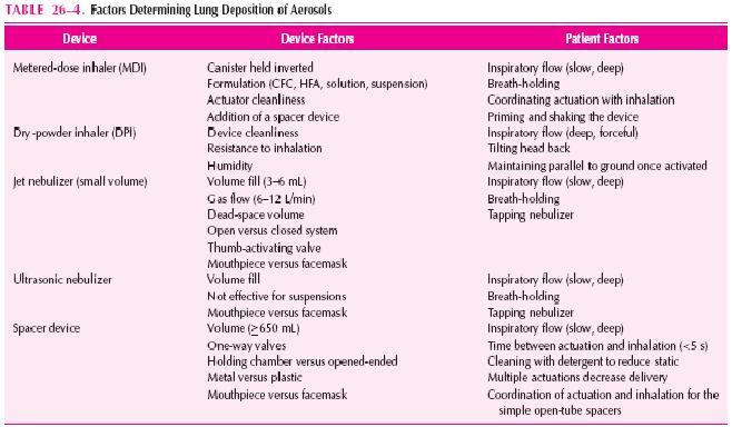
Aerosol delivery of drugs for asthma has the advantage of being site-specific and thus enhancing the therapeutic ratio.  Inhalation of short-acting β2-agonists provides more rapid bronchodilation than either parenteral or oral administration, as well as the greatest degree of protection against EIB and other challenges. Inhaled corticosteroids produce a greater reduction of BHR than corticosteroids administered systemically. Specific agents (e.g., cromolyn, nedocromil, formoterol, salmeterol, and ipratropium) are only effective by inhalation.  Given the international ban on the production and use of chlorofluorocarbons (CFCs), the manufacturers of CFC-propelled metered-dose inhalers (MDIs) are developing new devices for delivering topically active medication.  Therefore, an understanding of aerosol drug delivery is essential to optimal asthma therapy. Table 26-4 lists the factors determining lung deposition of therapeutic aerosols.

DEVICE DETERMINANTS OF DELIVERY
Devices used to generate therapeutic aerosols include jet nebulizers, ultrasonic nebulizers, MDIs, and dry-powder inhalers (DPIs).
 
The single most important device factor determining the site of aerosol deposition is particle size.  Devices for delivering therapeutic aerosols generate particles with aerodynamic diameters from 0.5 to 35 microns.  Particles larger than 10 microns deposit in the oropharynx, particles between 5 and 10 microns deposit in the trachea and large bronchi, particles 1 to 5 microns in size reach the lower
airways, and particles smaller than 0.5 microns act as a gas and are exhaled. In asthma, the airways, not the alveoli, are the target for delivery. Respirable particles are deposited in the airways by three mechanisms:
(1) inertial impaction,
(2) gravitational sedimentation, and
(3) Brownian diffusion. 
The first two mechanisms are the most important for therapeutic aerosols and probably are the only factors that can be manipulated by patients. Each delivery device within a classification generates specific aerosol characteristics, so extrapolation of delivery data from one device cannot be done for the other devices in the class. For instance, MDIs can deliver 5% to 50% of the actuated dose; DPIs, 10% to 30% of the labeled dose; and nebulizers, 2% to 15% of the starting dose.  MDIs and DPIs are portable and convenient, unlike nebulizers. MDIs consist of a pressurized canister with a metering valve; the canister contains active drug, low-vapor-pressure propellants such as chlorofluorocarbon (CFC) or hydrofluoroalkane (HFA), cosolvents, and/or surfactants.  With any change in these components, the Food and Drug Administration (FDA) considers it to be a new drug that requires stability, safety, and efficacy studies prior to approval. The drug is either in solution or a suspended micronized powder. In order to disperse the suspension for accurate delivery, the canister must
be shaken. The metering chamber measures a liquid volume, and therefore, the device must be held with the valve stem downward so that the chamber is covered with liquid  (Fig. 26-4). When the
canister is actuated, the device releases the propellant and drug in a forceful spray whose particles are large (mass median aerodynamic diameter [MMAD] = 45 microns)  (see Fig. 26–4). As evaporation
occurs, the particle size is reduced to a final MMAD of 0.5 to 5.5 microns depending on the MDI. The aerosol cloud of a CFCpropelled MDI extends at least 10 in beyond the MDI at the lowest
MMAD, and that of an HFA-propelled MDI extends about 6 in. The breath-actuated MDI Autohaler, is cocked with a lever to “load” the dose of medication, a baffle is opened by inspiratory pressure,
and the dose is expelled from the canister metering chamber. While the need for hand-lung coordination for proper actuation is reduced significantly with breath-actuated MDIs, these devices do not allow the use of a spacer device.

Spacer devices are used frequently with an MDI to decrease oropharyngeal deposition and enhance lung delivery. However, not all spacer devices produce similar effects. The design of spacers varies from simple open-ended tubes that separate the MDI from the mouth to holding chambers with one-way valves that open during inhalation (the preferred system). The purpose of a spacer is to allow evaporation of the propellant prior to inhalation. Use of a spacer allows inhalation after actuation of the device, obviating the need for good hand-lung coordination, and permits a greater number of drug particles to achieve a respirable droplet size.  Additionally, the largeparticles that normally would deposit in the oropharynx “rain out” in the spacer. All the available spacers significantly reduce oropharyngeal deposition from MDIs, with the holding-chamber devices being
superior to the open-ended tubes.  This reduction in oropharyngeal deposition is an important factor in reducing local adverse effects (i.e.,hoarseness and thrush) from inhaled corticosteroids.   The change in lung delivery depends on both the MDI and the drug, where one spacer device may enhance delivery with one MDI preparation and decrease delivery with others.  Finally, over time, holding chambers can build up static electricity that attracts small particles to the sides of the chamber, significantly reducing aerosol availability. It is recommended that spacers be washed weekly with household detergent, with a single rinse, and allowed to drip dry. Dry micronized powders can be inhaled directly into the lung. Four DPIs for asthma are available for use in the United States (i.e., Diskus, Rotahaler, Turbuhaler, and Aerolizer), with others under development.  Each has unique characteristics with advantages and disadvantages (Table 26–5). The primary advantage of DPIs is that they are breath-actuated and require minimal hand-lung coordination.  Some DPIs are more flow-dependent than others. Thus, similar to MDIs and spacers, delivery data from one DPI cannot
be extrapolated to another. Nebulizers come in two basic types, the jet nebulizer and the ultrasonic nebulizer. Jet nebulizers produce an aerosol from a liquid solution or suspension placed in a cup. A tube connected to a stream of compressed air or oxygen flows up through the bottom and draws the liquid up an adjacent open-ended tube. The air and liquid strike a baffle, creating a droplet cloud that is then inhaled.  Ultrasonic nebulizers produce an aerosol by vibrating liquid lying above a transducer
at speeds of about 1 mHz.  Both produce similar degrees of lung deposition, with the exception that ultrasonic nebulizers are ineffective for nebulizing micronized suspensions.  The aerosol output and
lung delivery vary significantly among the commercially available jet nebulizers even when operated in the same manner.  Increasing fill volume will increase the total amount of drug delivered; however, it
also will take longer for the patient to nebulize the dose. TheMMAD of the droplets is related directly to the gas flow, with flows of 5 to 12L/ min providing an aerosol cloud with an MMAD of 4 to 8 microns for most jet nebulizers.

PATIENT DETERMINANTS OF DELIVERY (See Table 26–4)
The most important patient factor determining aerosol deposition is inspiratory flow.  High inspiratory flows increase the degree of deposition owing to impaction of particles of any size, thereby increasing deposition centrally (i.e., throat and large airways) and decreasing peripheral deposition. Holding chambers enhance the clinical efficacy in patients with poor hand-lung coordination but may offer no advantage in patients who can use an MDI optimally alone. Optimal inspiratory flow for most MDIs is
slow and deep (approximately 30 L/min).  In general, DPIs require


higher inspiratory flows (≥60 L/min) and a change in inhalation technique (i.e., deep, forceful inspiration) for optimal actuation, which, in turn, increases the amount of drug delivered to the larger central airways.  However, this difference in delivery may not produce clinically significant differences.  Patients should be cautioned not to exhale into DPIs because this causes loss of dose and moistens the dry powder, causing aggregation into larger particles. Patient factors that cannot be controlled include interpatient variability in airway geometry (particularly the differences between children and adults)  and the effects of bronchospasm, edema, and mucus hypersecretion. Mild
obstruction increases aerosol deposition; however, severe obstruction probably leads to increased central deposition from impaction.  The absolute delivery to the lung is not as important as consistency of delivery, assuming that a sufficient dose to produce the desired therapeutic
effect is achieved. No single inhalation device is the best for all patients. Table 26–5 lists the differing characteristics of inhalation devices. Appropriate inhalation technique is essential to achieve optimal
drug delivery and therapeutic effect. The components are illustrated in Figure 26–5. Approximately 50% to 80% of a dose from MDIs and DPIs impacts on the oropharynx and is then swallowed;
the rest is either left in the device or exhaled.  It is important that actuation occurs during inhalation, although the time during inspiration is unimportant.  Although radiolabeled studies indicate improved
delivery by holding the actuator 2 to 3 cm in front of an open mouth to allow more evaporation and less impaction, physiologic studies with bronchodilators have failed to document an advantage for this
method.  Many patients do not use their MDIs optimally, and patient instruction with demonstration is the most effective means of improving inhaler technique. Even with instruction, up to 30% of
patients, particularly young children and the elderly, cannot master the use of an MDI. For these patients, attachment of a holding-chamber device to the MDI or use of a breath-actuated MDI can improve effi- cacy significantly.  Mouth rinsing following treatment with MDIand DPI-inhaled corticosteroids is important to minimize local effects and oral absorption. Delivery from high-resistance DPIs is more flow-dependent than from low-resistance DPIs. The Turbuhaler device has a greater flow dependency than the Aerolizer and the Diskus. Children 6 to 12 years of age received approximately one-half the dose of radiolabeled budesonide via Turbuhaler than those older than 12 years of age, whereas children younger than 6 years of age received

one-quarter of the dose. The Aerolizer and the Diskus deliver about 15% of the dose to the airways, and this appears to be similar at both 30 and 60 L/min inspiratory flows; both have been approved for children aged 5 and 4 years, respectively.
»»  READMORE...